Heel pain that is worst with the first steps in the morning is one of the most common reasons people look for custom insoles in Delhi NCR. Plantar fasciitis is a clinical diagnosis. A Materialise Footscan does not replace a doctor. What the scan can do is show how the foot is loading while you walk, so an insole can be designed around those loads instead of a generic arch shape.
At P.O.R.C in Safdarjung Enclave we use dynamic pressure mapping and PHITS 3D-printed shells, then add the top covers and local modifications the scan and the clinical exam both point to.
What the plantar fascia is doing
The plantar fascia is a thick band under the foot, from the heel to the forefoot. When the arch flattens too quickly, or the heel strikes with a high peak, that band can be overloaded. People often feel it as a stab or tightness under the heel, sometimes after standing all day in South Delhi traffic, on shop floors, or on hospital rounds.
Insoles will not “cure” that on their own. Stretching, footwear, load management and medical review still matter. The insole’s job is to change how pressure and motion are shared, so the fascia is not taking the same peak on every step.
What a dynamic 2D footscan can show
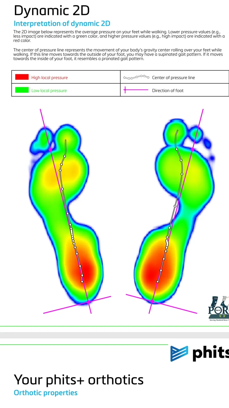
A dynamic scan records average pressure while you walk across the plate, not a still photograph of the foot. On the report, green is lower pressure. Red is a higher peak. A white dotted line is the centre of pressure: the path of body weight rolling from heel to toe.
If that line drifts toward the inside of the foot, the pattern is more pronated. If it stays toward the outside, it is more supinated. Many plantar-fascia irritations we see in clinic come with a red heel blob, a relatively unloaded midfoot, and a centre-of-pressure line that cuts in toward the first toe. That is a mechanical clue, not a label by itself.
Standing on the plate is only the start
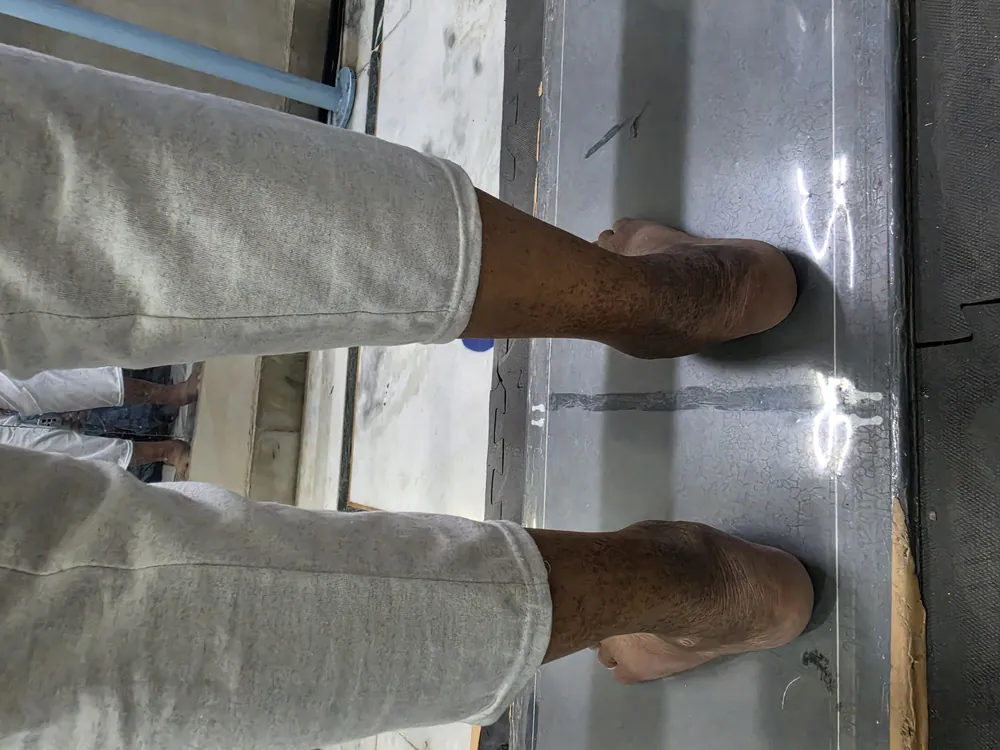
Before the software, we still look at the heels from behind. Inward roll of the heel (valgus) and a collapsing arch are things you can often see on the scanner platform. The Footscan then puts numbers and a pressure picture on that observation, using real-time dynamic data from that person’s gait.
How the custom insole is built
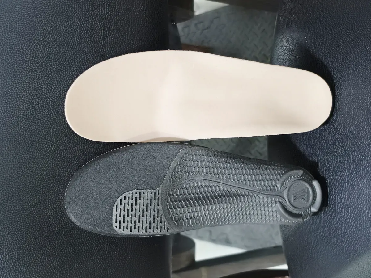
The shell we use most often for this work is a PHITS 3D-printed base, designed on the scan. The underside is not decoration. Honeycomb zones, grooves and local ridges are where the corrections live. On top of that shell we can add a soft Poron mid-layer and a Plastazote upper, so the foot is not sitting on a hard printed surface.
Typical modifications for a plantar-fascia pattern include the following. Not every pair needs every feature. Depth and position are set in millimetres on that person’s scan.
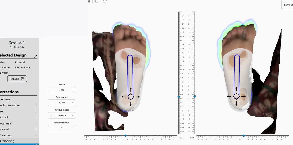
Plantar fascia groove
A longitudinal channel under the fascia so the band is not pressed into a hard ridge at the arch. In the design software this is an offloading tool: depth, width, length and a small rotation, placed along the scan rather than guessed from a shoe size. A common starting point is a few millimetres of depth and a groove that follows the fascia line, then we check comfort in the shoe.
First-metatarsal (1st ray) forefoot extension
A firmer or slightly extended zone under the first metatarsal head. That can help the big-toe side of the forefoot share push-off, instead of dumping load into the fascia at the end of stance. On the printed underside it often shows as a distinct ridged panel under the first ray.
Heel raise
A small lift under the heel reduces the stretch on the fascia at heel strike and in early stance. It is not a high shoe lift. It is a few millimetres, combined with a stable heel cup, when the scan shows a hot red heel and the person is sore first thing in the morning.
Navicular support
Support under the inner arch, near the navicular, so the arch does not collapse as far or as fast. That is the honeycomb region on many PHITS undersides. The aim is to slow pronation, not to jam a high rigid arch into every foot.
Soft Poron mid-layer and Plastazote upper
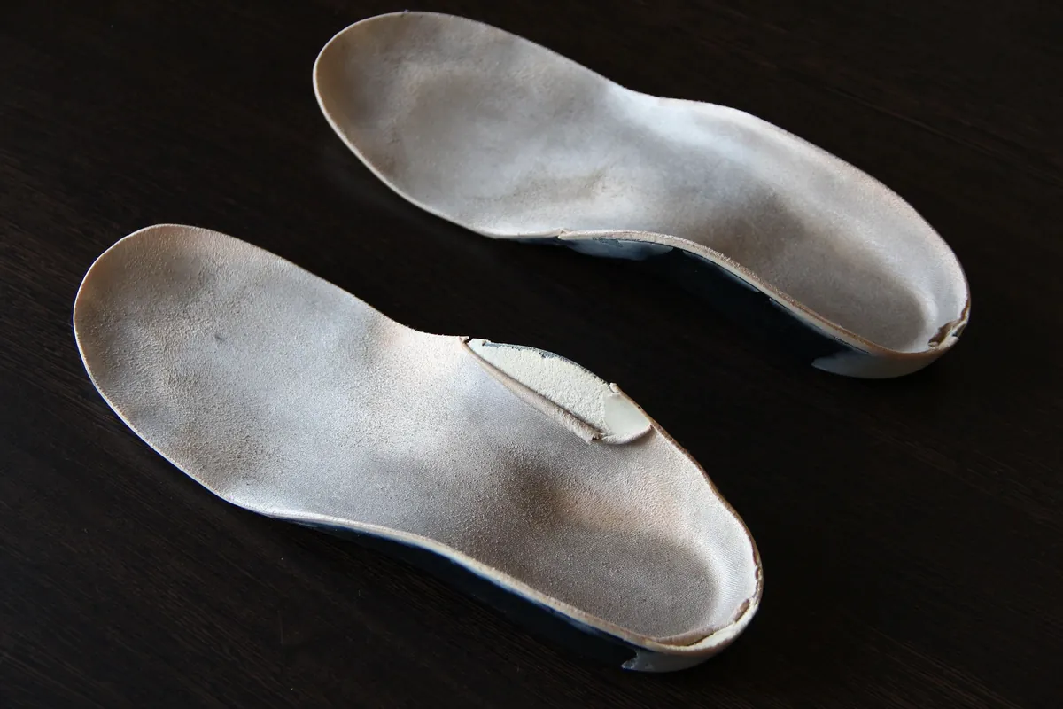
Poron is a shock-absorbing foam used as a thin mid-layer. Plastazote is a heat-formable polyethylene foam used as the top cover the skin actually meets. Together they take peak pressure off a printed shell, which matters for a sore heel and for anyone who will wear the pair all day. The beige top you see on a finished pair is usually that Plastazote cover.
What this is not
A red heel on a scan is not automatic proof of plantar fasciitis. A groove in an insole is not a substitute for ruling out stress fracture, nerve irritation or referred pain. If the heel is swollen, numb, or the pain is after an injury, get medical review first. We have written more on how P.O.R.C uses Footscan and PHITS for this problem here: Plantar fasciitis, Footscan and PHITS 3D insoles in Delhi.
Book a footscan in Delhi NCR
PHITS printing is done from our Safdarjung Enclave clinic. Assessment can start there, or you can call to confirm hours at the hospital branches.
- P.O.R.C, 42 Ground Floor, Street No. 1, Krishna Nagar, Safdarjung Enclave, New Delhi 110029
- P.O.R.C at Apollo Hospital, Sarita Vihar
- P.O.R.C at BLK-Max Super Speciality Hospital, Pusa Road
Call 9810006111 or WhatsApp 9811706111.